Aquatic Shops Near Me . Aquatic warehouse & nature's pets. Fish tanks, marine aquariums, pond pumps and fish supplies by birmingham based shirley aquatics. Visiting Chew Thean Yeang aquarium shop M're Undefined from blog.mohdimran.com Aquatic warehouse & nature's pets. Shop today and get free shipping on qualifying orders! At aquatic warehouse, we guarantee you will find something beautiful and unique for your own aquarium.
Get link
Facebook
X
Pinterest
Email
Other Apps
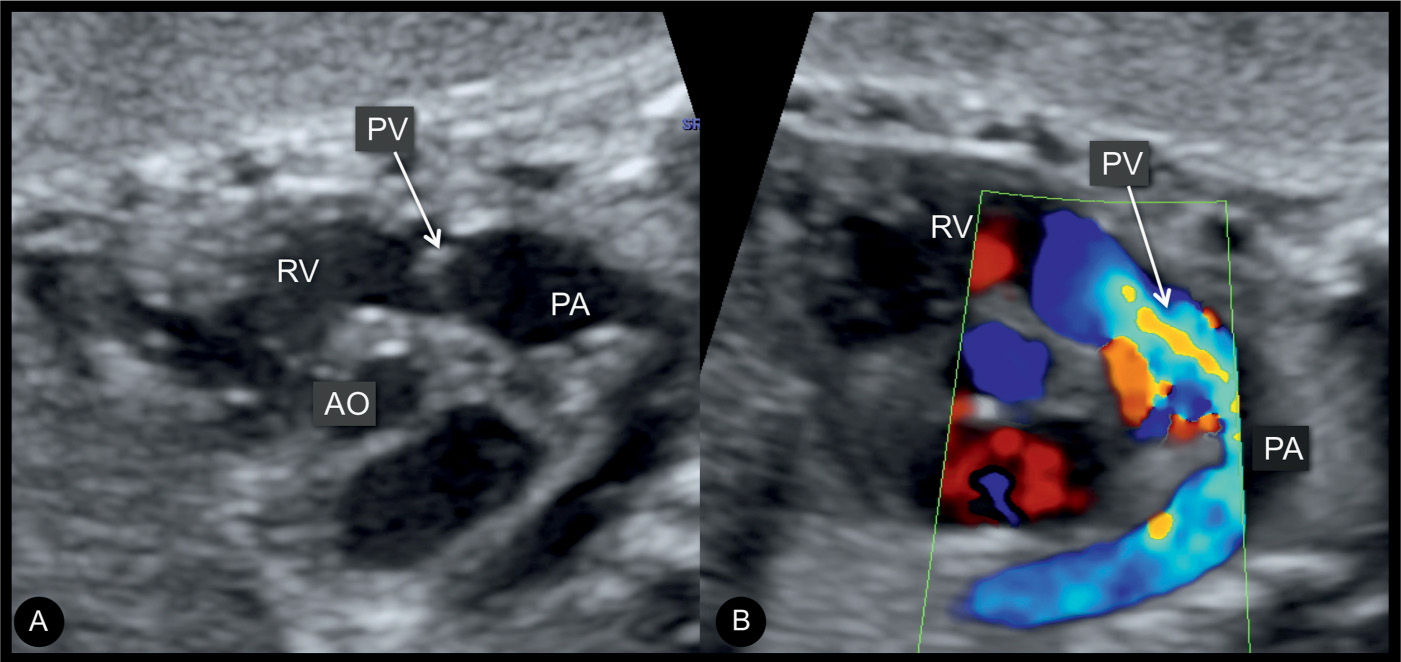
Ultrasound Pulmonary Artery Narrowing
Get link
Facebook
X
Pinterest
Email
Other Apps
-
Ultrasound Pulmonary Artery Narrowing. Pulmonary stenosis can be seen at the valvular level or at the infundibulum. To our knowledge, prenatal diagnosis of lpa sling has not been reported so far.
Pulmonary Stenosis, Pulmonary Atresia with Intact from obgynkey.com
Pulmonary stenosis can be seen at the valvular level or at the infundibulum. Pulmonary stenosis is obstruction of the right ventricular outflow tract caused primarily by narrowing of the infundibulum or valve stenosis. In chronic pulmonary thromboembolic disease, however, organization of the thrombus may result in no recognizable angiographic abnormalities or nonspecific findings.20 in addition, pulmonary
Thirty Patients Had Significant Pulmonary Artery Stenosis And Underwent Balloon Angioplasty.
This will give an estimate of the severity of obstruction. When narrowing is below the valve, this is called subvalvular pulmonary stenosis. Right ventricular hypertrophy (small rv chamber).
In All, Of 34 Branch Pulmonary Arteries Were Dilated.
To our knowledge, prenatal diagnosis of lpa sling has not been reported so far. This may be difficult to appreciate antenatally. Ultrasound assessment of anatomically determined stenosis of the vertical vein collecting pulmonary venous return and ultrasound diagnosis of pulmonary sling with proximal stenosis of left pulmonary artery and.
It Is Commonly Seen With Other Heart Defects.
Fourteen pregnant ewes were included in this study (gestational age, 90 to 120 days). The most common form is a dysplastic valve. We report a case in which lpa sling was diagnosed during fetal ultrasound examination.
Dilatation Of The Proximal Pulmonary Artery (Post Stenotic).
This study was designed to compare findings of ivus imaging and those of angiography of the pulmonary artery before and after the balloon angioplasty procedure. The valve of the pulmonary artery is made up of three thin leaflets, and its job is to control the flow of blood from the heart to the main pulmonary artery. The characteristic hemodynamic feature of pulmonary stenosis is an increased systolic pressure gradient between the pulmonary artery and the right ventricle 1,2.
Narrowing At The Valve Is Called Pulmonary Valve Stenosis.
Us contrast agent increases the intensity of the doppler. Right atrium may be dilated and have thickened walls. Intrauterine fetal doppler echocardiographic data obtained 15 days after surgery were compared with preoperative values.
Fontweight React Native Changes Fontstyle . Javascript answers related to “underline in react js”. Export default function app () { return ( fontstyle</strong>: React Native fontWeight 'bold', only shows in Android for from stackoverflow.com Javascript answers related to “how to change the color of word in a paragraph in react native”. React native font weight cheatsheet ios. Anytime one of them changes, updatefont is called which recreates the font from scratch.
Cn Patriotism Have Negative Impact On America . Too much nationalism may hurt international trade; The idea that someone can come to the us from anywhere and pursue their own version of happiness or achieve what they want to achieve in life is an effort many wish to have access to. Patriotism is not protesting a pandemic. Patriotism is from www.courant.com Patriotism may lead to unhuman behavior; Patriotism or nationalism can seek to divide people; Immigrants are a boon to the country’s economy.
Is It Possible To Get Negative Correlation And Positive R2 . If you’re correlating x with y and you run a regression predicting y from x, you should get exactly the same standardized beta weight as correlation (including the positive or. It depends on your research work but more then 50%, r2 value with low rmes value is acceptable to scientific research community, results with low r2 value of 25% to 30% are valid because. Rajandran R Blogs Nifty Futures Continues With Too Many from www.blogadda.com Note that the estimated slope of the line changes from a positive 179.5 to a negative 87.1 — just by removing one data point. To get that value you have to have zero error in your regression analysis. # of cigarettes/day dependent variable:


Comments
Post a Comment